Hossameldin Mohamed Selim Mohamed Selimpresents his thesis on 3D reconstruction of defects using a non-destructive test method based on laser-induced ultrasound
Jul 21, 2020
Mohamed Selim presents his thesis co-directed by Crina Cojocaru (Dept. of Physics) and Miguel Delgado tight (Dept. of Electronic Engineering), on July 21 at the Terrassa Campus. Titled "Hybrid senar-destructive technique for volumetric defect analysis and reconstruction by remote laser induced ultrasound"
This doctoral thesis deals with the design, study and implementation of a hybrid, non-contact method of non-destructive testing (NDT) for the analysis of metal objects containing internal defects or fractures. . We propose a hybrid opto-acoustic technique that combines ultrasound generated by laser impact as an exciter and ultrasonic transducers as receivers. The work proposes a detailed study of the detection and reconstruction in 1D, 2D, and 3D of defects present in a metal object, using the hybrid technique of NDT without contact and controlled remotely. Our device has several advantages over photonic and ultrasonic techniques, while reducing some disadvantages of these methods separately.
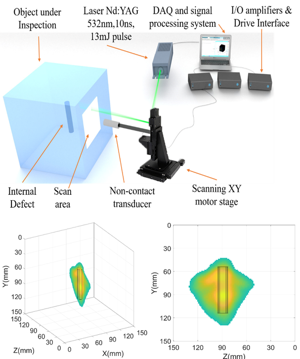
Our method combines experimental results with numerical simulations based on high-resolution signal processing. The experimental setup consists of a pulsed laser of ns at a wavelength of 532 nm, which impacts the surface of the object. The laser pulse is absorbed, creating a localized thermoelastic expansion that induces a broadband ultrasonic pulse that propagates through the material. The remote-controlled laser scans a selected area of the object's surface. For each excitation point, the ultrasound propagates through the object and is reflected or dispersed to the defects of the material. These ultrasound waves are detected by transducers and recorded in a data acquisition system that will be processed later. In a first step, by analyzing the flight time, we can locate and determine the size of the defect in a 1D view. The capabilities of detecting internal defects in a metal sample are also studied by wavelet transformation due to their multi-resolution characteristics in time and frequency.
The details of the defects are recorded from different angles, achieving a complete 3D reconstruction. Finally, we show our results in a complementary topic, related to a particular case of ultrasonic propagation in solids. From the beginning, we wanted to have a physical understanding of the propagation and diffraction of ultrasound waves in solid materials. Controlling diffraction patterns in solids, through the use of ultrasonic lenses, would help focus / collimate the ultrasound, reducing echoes and reflections on the contour surface, and improving the NDT analysis process.
A new spatial clustering algorithm is applied and the resulting time and frequency maps are used. Our method combines experimental results with numerical simulations based on high-resolution signal processing. The experimental setup consists of a pulsed laser of ns at a wavelength of 532 nm, which impacts the surface of the object. The laser pulse is absorbed, creating a localized thermoelastic expansion that induces a broadband ultrasonic pulse that propagates through the material. The remote-controlled laser scans a selected area of the object's surface. For each excitation point, the ultrasound propagates through the object and is reflected or dispersed to the defects of the material. These ultrasound waves are detected by transducers and recorded in a data acquisition system that will be processed later. In a first step, by analyzing the flight time, we can locate and determine the size of the defect in a 1D view. The capabilities of detecting internal defects in a metal sample are also studied by wavelet transformation for their multi-resolution
Phonic crystals are used to regulate the diffraction and frequency response of ultrasonic waves that propagate in fluids. However, these structures have been studied much less in solid materials. We performed detailed numerical simulations of ultrasonic propagation on a solid phonic crystal and demonstrated focusing and autoclimation effects.
We finally attached our phonic crystal lens to the solid object under study, demonstrating that diffraction control is conserved inside this object using the coupling material.
characteristics in time and frequency.ia to estimate the position of the defect. For the 2D visualization of the defects, we extend the analysis of the signal using the synthetic aperture focusing technique (SAFT). We implemented a new 2D apodization filter, along with the SAFT technique, and demonstrated that it eliminates unwanted effects, improving the resolution of the reconstructed image of the defect. The next step is a 3D analysis and reconstruction. In this case we achieve a fully automated and contactless experimental configuration, allowing sweeping areas on different faces of an object.

Share: